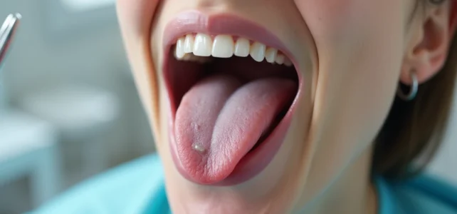
A small whitish area on the edge of the tongue, a sore that refuses to heal for three weeks: one often hesitates between a common canker sore and something more concerning. Tongue cancer, primarily a squamous cell carcinoma, is strongly linked to tobacco and alcohol, but also affects younger profiles through human papillomavirus (HPV). Knowing how to distinguish a suspicious lesion from a temporary irritation allows for timely consultation.
Leukoplakia on the tongue: when a white spot becomes suspicious
On the ground, the most common situation is this: a white patch is noticed on the lateral edge of the tongue, sometimes slightly rough to the touch. One thinks of food deposits, a burn, or dental irritation. We wait. And it is precisely this delay that poses a problem.
This white spot is called leukoplakia. It corresponds to an abnormal thickening of the oral mucosa. Not all leukoplakias are cancerous, but some represent a precancerous condition that can progress to squamous cell carcinoma if left untreated.
The distinguishing criterion is not the color itself, but the persistence. A classic canker sore disappears within about ten days. A precancerous leukoplakia, however, does not change despite mouth rinses and local treatments. Consulting photos of tongue cancer and white spots helps visualize the difference between a trivial irritation and a lesion that warrants a quick medical appointment.
Another point to observe: texture. A homogeneous and smooth surface is less concerning than an irregular plaque, mixing white and red areas (this is referred to as erythroleukoplakia). This mixed form presents a higher risk of transformation.

Persistent sore and unilateral pain: concrete signals to watch for
Leukoplakia is not the only signal. Several symptoms, taken in isolation, seem benign. It is their combination and duration that should raise concern.
- An ulcer that does not heal after three weeks, despite local treatment. It is often located on the side of the tongue, sometimes under the tongue, rarely in the center.
- Unilateral pain radiating to the ear, with no identifiable ENT cause. This type of referred pain is characteristic of lesions in the mobile part of the tongue.
- A palpable, hard, painless cervical lymph node, recently appeared on one side of the neck. Its presence may indicate local tumor extension.
- Discomfort when swallowing or speaking, with a persistent sensation of a foreign body in the mouth.
There is a tendency to rationalize each symptom separately. Ear pain becomes an ear infection, the lymph node a simple cold, the sore a dental trauma. The reflex to adopt: any oral lesion that persists beyond three weeks requires medical advice, whether it is painful or not.
Base of the tongue: more discreet symptoms
Tumors at the base of the tongue, often linked to HPV 16, pose a particular problem. This area, located at the back, is not visible in a mirror. The first signs often limit themselves to difficulty swallowing or a changed voice. Diagnosis frequently occurs at a more advanced stage, even though these HPV-related cancers generally respond better to radiotherapy and chemotherapy.
Risk factors for tongue cancer: tobacco, alcohol, and HPV
One cannot discuss visual recognition without addressing what concretely increases the risk. Three factors dominate, and their combination significantly multiplies the probabilities.
Tobacco remains the primary risk factor. Smoking exposes the oral mucosa to direct carcinogenic agents, promoting the appearance of precancerous lesions like leukoplakia. Alcohol, consumed regularly, acts as a solvent that facilitates the penetration of these toxic substances into the tissues.
The combination of tobacco and alcohol does not merely add risks, it multiplies them. This is a point often underestimated by patients who consume both moderately.
The third factor changes the usual patient profile. HPV, particularly strain 16, is now recognized as responsible for an increasing share of cancers of the base of the tongue and oropharynx. These cases affect younger patients, sometimes non-smokers and non-drinkers. Vaccination against HPV, recommended in France for both boys and girls, currently represents the best prevention against these forms of tongue cancer.

Diagnosis and biopsy: what happens during a consultation
When presenting with a suspicious lesion, the doctor (general practitioner, dentist, or ENT specialist) first conducts a clinical examination of the oral cavity. They palpate the tongue, the floor of the mouth, and the cervical lymph nodes. This initial examination is sometimes sufficient to strongly guide the diagnosis.
If the lesion is deemed suspicious, a biopsy is performed: a sample of tissue is taken and analyzed in pathology. This is the only examination that formally confirms the cancerous nature of a lesion. Neither a photo nor a visual examination alone can diagnose tongue cancer.
Depending on the results, an extension assessment (imaging, CT scan, MRI) specifies the stage of the tumor. This stage conditions the treatment: surgery alone for early stages, a combination of surgery and radiotherapy for more advanced stages, sometimes associated with chemotherapy.
An oral brushing test under evaluation
Recent studies are exploring an oral brushing test capable of identifying abnormal cells in the oral cavity. This type of screening, less invasive than a biopsy, could ultimately facilitate early detection, but it remains in the evaluation phase and does not yet replace the clinical examination followed by a biopsy.
Detected at an early stage, tongue cancer is treated with a significantly more favorable prognosis. A persistent white spot, a sore that does not heal, a new cervical lymph node: three situations that justify making an appointment without delay. The dentist, often the first professional to observe the oral cavity, plays a sentinel role that is too little exploited in the screening of these lesions.